·论 著·
李 勇,赵新鸿,孟晓东,陈 涛,刘 健,邱建宏*
(中国人民解放军白求恩国际和平医院泌尿外科,河北 石家庄 050082)
[摘要] 目的探讨磷脂酰肌醇蛋白聚糖 3(Glypican-3,GPC-3)蛋白在前列腺癌组织中的表达及意义。方法选取前列腺癌和前列腺增生组织各30例标本,采用免疫组织化学SP法染色,分析GPC-3蛋白在前列腺癌及前列腺增生组织中的表达情况。 结果GPC-3蛋白在前列腺癌组织中的阳性表达率高于前列腺增生组织(P<0.05),Gleason评分≥7分的前列腺癌组织中GPC-3蛋白的阳性表达率高于Gleason评分<7分者(P<0.05)。结论GPC-3蛋白在前列腺癌组织中高表达,可能与前列腺癌的发生、发展有关。
[关键词] 前列腺肿瘤;前列腺增生;磷脂酰肌醇蛋白聚糖类
doi:10.3969/j.issn.1007-3205.2016.04.007
随着人口老龄化、饮食结构和生活习惯的改变,我国前列腺癌的发病率和病死率呈逐年上升的趋势,已成为严重影响我国老年男性身体健康的疾病之一[1]。目前对于前列腺癌标志物的研究进展相对缓慢,无法满足临床需求,因此寻求更多有效辅助前列腺癌诊断的肿瘤标志物意义重大。磷脂酰肌醇蛋白聚糖 3(Glypican-3,GPC-3)定位于人类染色体X26.1,基因组结构全长大于900 kb,可编码580个氨基酸的蛋白质,相对分子质量约为66 000[2]。GPC-3是硫酸乙酰肝素糖蛋白家族的一员[3]。目前认为 GPC-3的HS链与生长因子及其受体、细胞外基质蛋白等大量生物效应分子相互作用,参与调节细胞形态和各种细胞行为(黏附、增殖、迁移和分化等)[4-5]。研究发现GPC-3基因的表达变异与多种肿瘤的发生有关,尤其对原发性肝癌的诊断具有重要意义,其特异性高于甲胎蛋白[6]。本研究采用免疫组织化学技术检测GPC-3蛋白在前列腺癌和前列腺增生组织中的表达,旨在分析GPC-3蛋白能否成为辅助诊断前列腺癌的肿瘤标志物。现报告如下。
1.1 一般资料 选取1998年8月—2012年6月中国人民解放军白求恩国际和平医院泌尿外科前列腺癌和前列腺增生组织各30例标本。前列腺癌组30例,年龄56~89岁,平均(71.0±7.8)岁;前列腺体积13.89~192.20 mL,平均(73.3±18.3) mL;血清总前列腺特异性抗原(prostate specific antigen,PSA)6.6~177.0 μg/L,平均(51.8±12.4)μg/L; Gleason评分≥7分26例,Gleason评分<7分4例;临床分期Ⅰ~Ⅱ期22例,Ⅲ~Ⅳ期 8例。前列腺增生组30例,年龄43~79岁,平均(69.8±6.2)岁;前列腺体积13.84~189.20 mL,平均(75.1±22.3) mL;血清PSA 1.8~15.3 μg/L,平均(4.3±1.7) μg/L。2组年龄、前列腺体积比较差异均无统计学意义(P>0.05),具有可比性。
1.2 方法 石蜡标本4 μm切片连续切片,采用免疫组织化学SP法进行染色,同时设置阳性对照和阴性对照,以试剂盒自带的阳性片免疫组织化学染色结果为阳性对照,以磷酸缓冲盐替代一抗的免疫组织化学染色结果为阴性对照。光镜下观察GPC-3的表达情况。
1.3 结果判定 采用半定量计分法判定,选取5个高倍镜视野(×400),按切片中阳性细胞比例分别记0~4分:阳性细胞≤5%为0分,>5%~25%为1分,>25%~50%为2分,>50%~75%为3分,>75%为4分;按染色深浅分别记0~3分,染色深度以多数细胞为准,无阳性细胞为0分,黄色为1分,棕黄色为2分,棕褐色为3分;阳性细胞的百分率得分与免疫组织化学染色强度的得分的乘积分成4个等级:0分为(-),1~ 2分为(+),3~ 4分为(++),5~ 12分为(+++),最终(-)和(+)记为低表达,(++)和(+++)记为高表达[7]。
1.4 统计学方法 应用SPSS 13.0统计学软件进行数据分析,计数资料以百分率表示,组间比较采用χ2检验。P<0.05为差异有统计学意义。
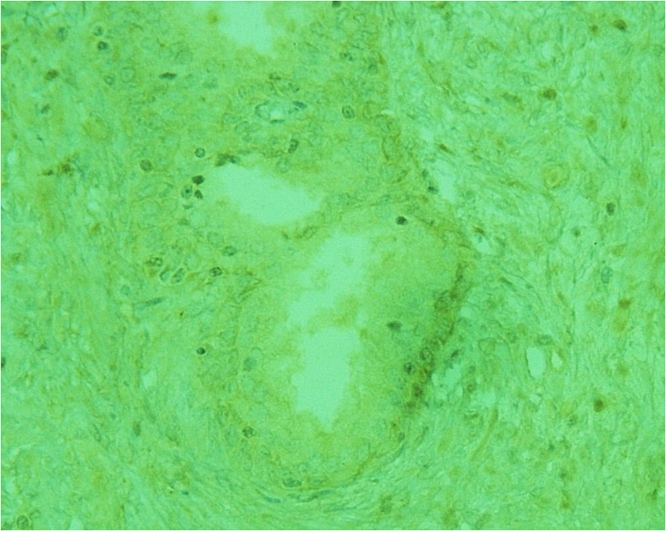
GPC-3蛋白在前列腺增生组织中阳性表达较少,少量细胞的细胞质着色,染色浅,呈黄色或浅黄色颗粒(图1);GPC-3蛋白在前列腺癌组织中阳性表达较多,大量细胞的细胞质着色,染色较深,呈深黄色或棕褐色颗粒(图2)。
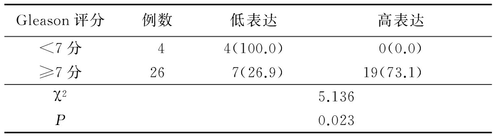
GPC-3蛋白在前列腺癌组织中的高表达率高于前列腺增生组织,差异有统计学意义(P<0.05);GPC-3蛋白在Gleason评分≥7分前列腺癌组织中的高表达率高于Gleason评分<7分者,差异有统计学意义(P<0.05)。见表1, 2。
图1 GPC-3蛋白在前列腺增生组织中的表达(免疫组织化学 ×400)
Figure 1 Expression of GPC-3 in benign prostatic hyperplasia tissue(immunohistochemistry ×400)
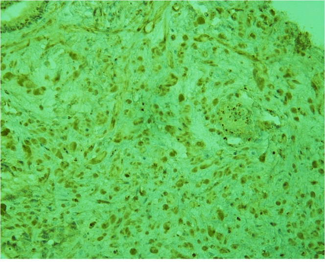
图2 GPC-3蛋白在前列腺癌组织中的表达(免疫组织化学 ×400)
Figure 2 Expression of GPC-3 in prostate cancer tissue(immunohistochemistry ×400)
表1 GPC-3蛋白在前列腺增生和前列腺癌组织中的表达
Table 1 Expression of GPC-3 in prostate cancer and benign prostatic hyperplasia (n=30,例数,%)
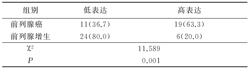
表2 不同Gleason评分前列腺癌组织中
GPC-3蛋白的表达
Table 2 Expression of GPC-3 in prostate cancer with different gleason score (例数,%)
目前,前列腺癌的早期诊断和治疗仍是泌尿外科学者们研究的热点。本研究采用免疫组织化学方法检测GPC-3蛋白在前列腺癌和前列腺增生组织中的表达情况,探索GPC-3蛋白成为前列腺癌肿瘤标志物的可能性。
文献报道,GPC-3蛋白在肝癌等肿瘤组织中高表达,与肝癌等肿瘤的关系密切[8]。Toretsky等[9]认为GPC-3可以作为辅助诊断肝癌的肿瘤标志物。Song等[10]研究认为在肝癌细胞中GPC-3蛋白通过自分泌和旁分泌的方式激活经典或非经典Wnt信号通路,刺激Wnt基因表达。Wnt基因产物具有促进组织细胞增殖及恶变的作用[11]。本研究结果显示,GPC-3蛋白在前列腺癌组织中的阳性表达率明显高于前列腺增生组织。说明 GPC-3蛋白在前列腺组织中高表达时,组织癌变可能性较大,GPC-3蛋白与前列腺癌的发生有关。
Takahashi等[12]认为Wnt信号通路的异常激活可诱导前列腺癌的发生、发展。Peng 等[13]用免疫组织化学和计算机图像分析检测肝细胞癌组织标本中GPC-3基因表达情况,结果发现肝细胞癌组织中GPC-3基因表达水平随着肿瘤分化程度的降低而升高。Capurro 等[14]发现Wnt基因产物可促进Wnt信号通路与其特异受体结合形成复合物,从而刺激肿瘤组织增殖和导致肿瘤细胞恶性程度升高并持续增殖。而且Wnt基因产物可以刺激核转录因子κB途径,能够与有些基因上的启动子结合,从而启动基因转录[15-16]。本研究结果显示,GPC-3蛋白在不同分化程度的前列腺癌组织中阳性表达率不同,前列腺癌细胞分化程度越差,阳性表达率越高。说明GPC-3蛋白高表达可能与前列腺癌的分化、恶性程度有关[17]。
[参考文献]
[1]韩苏军,张思维,陈万青,等.中国前列腺癌发病现状和流行趋势分析[J].临床肿瘤杂志,2013,18(4):330-334.
[2]Nakatsura T,Komori H,Kubo T,et al. Mouse homologue of a novel human oncofetal antigen,glypican-3,evokes T-cell-mediated tumor rejection without autoimmune reactions in mice[J]. Clin Cancer Res,2004,10(24):8630-8640.
[3]Pilia G,Hughes-Benzie RM,MacKenzie A,et al. Mutations in GPC3,a glypican gene,cause the Simpson-Golabi-Behmelover growth syndrome[J]. Nat Genet,1996,12(3):241-247.
[4]Seidler HB,Utsuyama M,Nagaoka S,et al. Expression level of Wnt signaling components possibly influences the biological behavior of colorectal cancer in different age groups[J]. Exp Mol Pathol,2004,76(3):224-233.
[5]Nelson WJ,Nusse R. Convergenece of Wnt,beta -catenin,and eadherin pathways[J]. Science,2004,303(5663):1483-1487.
[6]Sung YK,Hwang SY,Park MK,et al. Glypican-3 is overexpressed in human hepatoeellular carcinoma[J]. Cancer Sci,2003,94(3):259-262.
[7]邢传平,刘斌,董亮.免疫组织化学标记结果的判断方法[J].中华病理学杂志,2010,30(4):318.
[8]Weksberg R,Squire JA,Templeton DM. Glypicans:a growing trend[J]. Nat Genet,1996,12(3):225-227.
[9]Toretsky JA,Zitomersky NL,Eskenazi AE,et al. Glypican-3 exppssion in Wilms tumor and hepatoblastoma[J]. J Pediatr Hematol Oncol,2001,23(8):496-499.
[10]Song HH,Shi W,Xiang Y,et al. The loss of glypican-3 induces alterations in Wnt signaling[J]. J Biol Chem,2005,280(3):2116-2125.
[11]Capurro MI,Xiang YY,Lobe C,et al. Glypican-3 promotes the growth of hepatocellular carcinoma by stimulating canonical Wnt signaling[J]. Cancer Res,2005,65(14):6245-6254.
[12]Takahashi S,Watanabe T,Okada M,et al. Noncanonical Wnt signaling mediates androgen-dependent tumor growth in a mouse model of prostate cancer[J].Proc Natl Acad Sci U S A,2011,108(12):4938-4943.
[13]Peng F,Wu Z,Li X. Relationship between glypican-3 and Notch1 expressions in hepatocellular carcinoma[J]. Nan Fang Yi Ke Da Xue Xue Bao,2013,33(4):590-592,597.
[14]Capurro M,Martin T,Shi W,et al. Glypican-3 binds to Frizzled and plays a direct role in the stimulation of canonical Wnt signaling[J]. J Cell Sci,2014,127(Pt 7):1565-1575.
[15]Lustig B,Behrens J. The Wnt signaling pathway and its role in tumor development[J]. Cancer Res Clin Oncol,2003,129(4):199-221.
[16]De Cat B,Muyldermans SY,Coomans C,et a. Processing by proprotein convertases is required for glypican-3 modulation of cell survival,Wnt signaling,and gastrnlation movements[J]. J Cell Biol,2003,163(3):625-635.
[17]Filmus J,Capurro M. The role of glypican-3 in the regulation of body size and cancer[J]. Cell Cycle,2008,7(18):2787-2790.
(本文编辑:赵丽洁)
LI Yong, ZHAO Xin-hong, MENG Xiao-dong, CHEN Tao, Liu Jian, QIU Jian-hong*
(Department of Urology, Bethune International Peace Hospital, Shijiazhuang 050082, China)
[Abstract] Objective To investigate the expression and significance of glypican-3(GPC-3) in prostate cancer. Methods A total of 30 cases of prostate cancer tissues and 30 cases of benign prostatic hyperplasia tissues were included in the study with the immunohistochemical S-P method to detect the expression of GPC-3 in prostate cancer and benign prostatic hyperplasia. Results The positive rates of GPC-3 expression in prostate cancer tissues was significantly higher than that in benign prostatic hyperplasia tissues(P<0.05). The positive rates of GPC-3 expression in prostate cancer tissues with gleason score equal to or more than 7 was significantly higher than that with gleason score less than 7(P<0.05). Conclusion High expression of GPC-3 in prostate cancer may play an important role in development of prostate cancer.
[Key words] prostatic neoplasms; prostatic hyperplasia; glypicans
[收稿日期]2015-03-06;[修回日期]2016-02-03
[基金项目]河北省医学科学研究重点课题(20150370)
[作者简介]李勇(1985-),男,黑龙江绥化人,黑龙江省绥化市第一医院医师,医学硕士,从事泌尿外科疾病诊治研究。
[中图分类号] R737.25
[文献标志码] A
[文章编号] 1007-3205(2016)04-0397-03
*通讯作者。E-mail:hpyyqiujh@163.com